

Pictures of IVF und ICSI
Ultrasound-guided transvaginal egg collection in the woman under ultra-short anaesthesia.
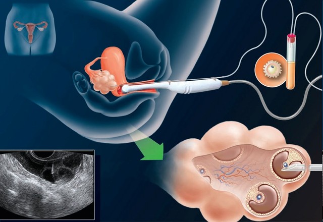
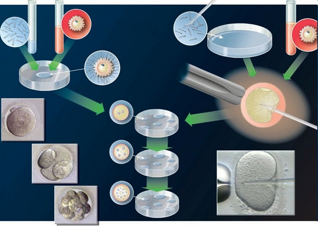
Conventional IVF (left) and microinjection (ICSI, right).
Ultrasound-guided embryo transfer.
![]()
Pictures from the IVF laboratory
The following pictures about the first moments of human life possess an unadorned aesthetics that hardly anyone can escape.
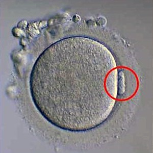
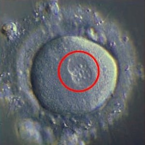
The human germ cells (gametes): a mature egg on the upper left which has been aspirated from the ovary (the circled polar body indicates that the egg is mature), on the right a zygote (fertilised egg) the next morning, exhibiting two bubbles (the so-called pronuclei with male and female chromosomes) ready to merge. During this fusion (called syngamy), male and female genetic material combine at random into a new human being.
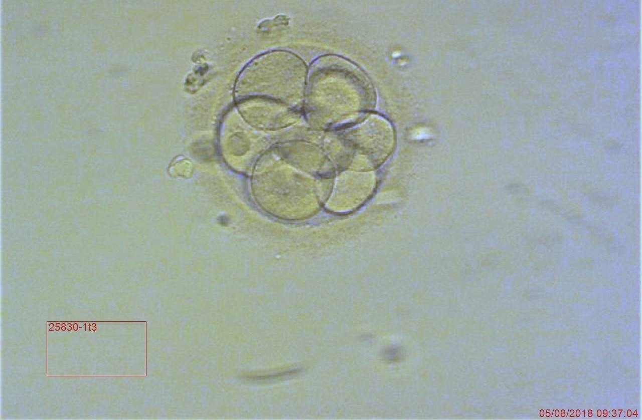
A ten-cell embryo on day 3 with optimal optical quality. Replacement of the embryo(s) in the womb (the embryo transfer) will take place on day 2, 3 or 5.
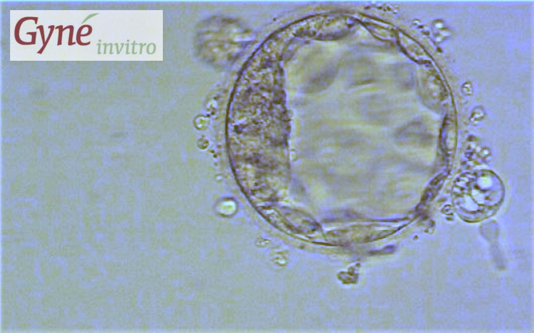
On the fifth day, a cavity has formed inside the embryo. This tiny hollow sphere is called blastocyst. A first specialisation of the cells has taken place - the darker inner cell mass on the left will form the embryo, the outer layer (trophectoderm) the placenta. The baby from precisely this blastocyst will be born in November 2018!